What is macular degeneration?

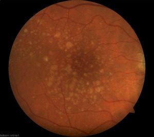
What a retina with dry macular degeneration looks like.
In Austin, TX, Macular Degeneration, also known as AMD and ARMD (for age related macular degeneration), is a leading cause of visual loss in older adults. The exact cause of this disease is unknown but is likely related to a variety of factors including genetic predisposition and inflammation. Visual loss results from damage to the central portion of the retina called the macula. The macula is responsible for the sharp and direct vision needed for activities such as reading, driving or recognizing fine details. This disease is commonly related to aging and is most prominent in patients over 65 years old.
Types of macular degeneration
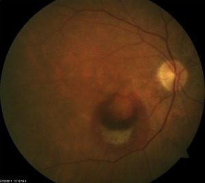
Macular degeneration is often grouped into two categories. These are the dry and wet types of macular degeneration. The dry form is commonly found earlier in the disease course. The changes include loss of retinal pigment and deposit of material in the tissues underlying the macula. These changes can lead to decreased or distorted vision. The progression of dry macular degeneration is not typically rapid. As the disease progresses changes in the blood vessels underlying the retina can cause leaking or bleeding which is the “wet” component of wet macular degeneration.
Evaluation

What a retina with wet macular degeneration looks like.
Our Austin patients with dry macular degeneration are encouraged to self-monitor their vision. This is done with the use of an amsler grid such as seen to the side. The grid is held at reading distance from the eye. Cover each eye in turn and look at the central spot with the uncovered eye. Normally the grid would appear as straight lines. In eyes with changes from AMD the lines may be distorted or blank spaces where the lines are not visible may be present. It is also possible to monitor by looking at an object with straight lines such as window blinds. If the lines have new changes including distortion or waviness it is possible that the AMD has progressed.
During your office visit in our Austin location, an evaluation with one of the country’s premier retina specialists, Dr. Stephen Smith, will be completed with an exam and diagnostic studies including photographs, dye studies (fluorescein angiogram) for leakage and mapping of the macula area with computerized imaging (optical coherence tomography or OCT).
Treatment
There are no treatments for the dry form of AMD. However, it is important to monitor your vision for possible progression. Use of an amsler grid on an ongoing basis is encouraged to detect early progression to the wet form of macular degeneration. Patients may also be asked to begin vitamin supplementation and to add green, leafy vegetables and fish oil to their diets.
Today’s treatment for the wet form of macular degeneration most commonly involves injection of medication within the eye. The two medicines most used are avastin and lucentis. Both medicines act by blocking a hormone that contributes to vascular leakage. By blocking the hormone and reducing the impetus to leakage or bleeding, the eye has a chance of drying the macular area and restoring macular anatomy and allowing improved vision.
